PLANT CELL ATLAS
  • Home
  • About
    • People
    • Motivation
    • Goals
    • Milestones
    • Affilations
    • News >
      • Breaking News
      • Past News
    • Code of Conduct
    • Bylaws
  • Events
    • Upcoming Events
    • Previous Events
  • Resources
    • 2023 PCA Travel Award >
      • 2023 Awardee
      • 2022 Awardees
    • Job Posts
    • Fellowships
    • Publications >
      • 2022 Plant Physiology Focus Issue
    • PCA Videos
    • Annual Report
  • Outreach
    • JR Biotek-PCA Best Talk Winner
    • PCA Art Exhibit >
      • About
      • Plant Biotechnology
      • Black Plant Scientists
      • Bowie State University
      • Howard University
      • Morgan State University
    • Futurum Brochure
  • Contact
Plant Cell Atlas Spatial Metabolomics ​Workshop
Virtual Workshop
October 19, 2021


Objectives

Registration

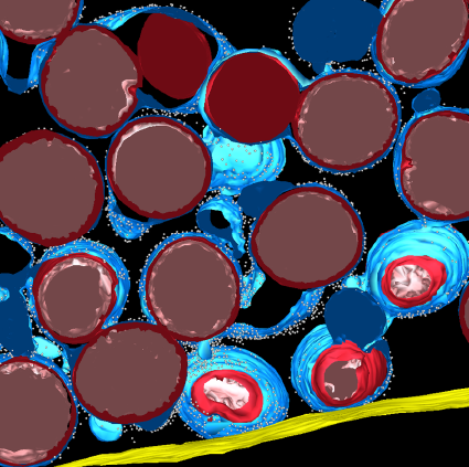
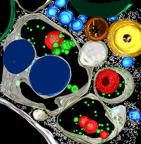
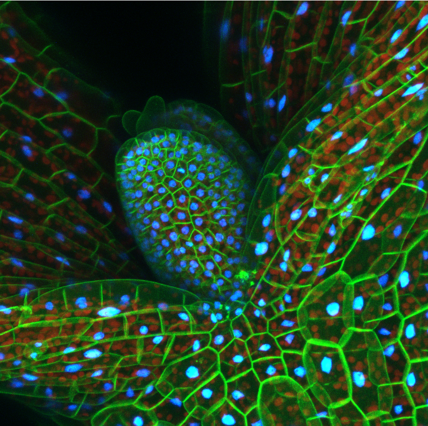
The goal of the Plant Cell Atlas (PCA) is to bring together a community who will comprehensively describe plant cell types by integrating high-resolution subcellular and cellular location information of nucleic acids, proteins, and metabolites. In line with achieving this goal, metabolomics provides a functional readout of the cellular and molecular programs controlled through expression of genes and proteins, as metabolites are directly linked to cellular function, response to environmental stresses, and progression to disease. Significant advances in mass spectrometry imaging and spatially resolved mass spectrometry technologies now make high-spatial resolution metabolomics notably more accessible for plant science. This PCA workshop aims to bring together world leaders in spatial and mass spectrometry imaging-based metabolomics from plant sciences and beyond to help realize the development of a comprehensive PCA metabolome.
Registration is closed for the PCA Spatial Metabolomics Workshop.

Workshop Times
PDT - 8:00am - 11:00am
MDT - 9:00am - 12:00pm
CDT - 10:00am - 1:00pm
EDT - 11:00am - 2:00pm
CEST - 5:00pm - 8:00pm
GMT+8 - 11:00pm - 2:00am (10/20/2021)

Recordings of the most of the talks will be posted to the PCA YouTube channel in mid-November.

Workshop Organizers and Speakers

Epidermal plasma membrane in green and nuclei (H2B-TFP) in blue, together with chlorophyll autofluorescence in red below in the mesophyll. Photo credit: Kate Harline (Roeder Lab)
Pin1-YFP (yellow) and DR5::RFP (red) in the maize shoot apex. Photo credit: Leiboff Lab
Depth-coded image of microtubules in root hairs of the model grass, Brachypodium distachyon. Photo credit: Elison Blancaflor
 

Agenda

8:00 AM - 8:05 AM PDT
Introduction
8:05 AM - 8:30 AM PDT
Akos Vertes (George Washington University)
Talk Title: Basic concepts in single cell analysis and spatial metabolomics of plant tissues
8:30 AM - 8:55 AM PDT
Theodore Alexandrov (European Molecular Biology Laboratory - EMBL)
Talk Title: METASPACE and SpaceM, tools for spatial single-cell metabolomics
8:55 AM - 9:20 AM PDT
Hua Tian (Pennsylvania State University)
Talk Title: Successive high-resolution (H2O)n-GCIB and C60-SIMS imaging integrates spatial omics (metabolites, lipids and proteins) in single cells on frozen-hydrated tissues
9:20 AM - 9:35 AM PDT
Break
9:35 AM - 10:00 AM PDT
Ingela Lanekoff (Uppsala University)
Talk Title: Quantitative spatial mapping and in-depth characterization of metabolites and lipids
10:00 AM - 10:25 AM PDT
Young-Jin Lee (Iowa State University)
Talk Title:
High-spatial resolution mass spectrometry imaging of plant metabolites with matrix-assisted laser desorption ionization
10:25 AM - 11:00 AM PDT
Panel Discussion: Future outlook for single cell metabolomics in plants
Protein bodies in maize endosperm, Marisa Otegui
Electron tomographic reconstruction of protein bodies in maize endosperm. Photo Credit: Marisa Otegui
Arabidopsis cotyledon, Flavia Bossi
Reconstruction of an Arabidopsis first leaf from a six-day old seedling. Photo Credit: Flavia Bossi
Developing maize aleurone cell, Marisa Otegui
Electron tomographic reconstruction of a developing maize aleurone cell. Photo Credit: Marisa Otegui
STANFORD LAND ACKNOWLEDGEMENT
“The Plant Cell Atlas operates predominantly out of the Carnegie Institution for Science's Plant Biology Department located at Stanford University. Stanford sits on the ancestral land of the Muwekma Ohlone Tribe. This land was and continues to be of great importance to the Ohlone people. Consistent with our values of community and inclusion, we have a responsibility to acknowledge, honor, and make visible the University’s relationship to Native peoples.”

    stay up to date on Plant Cell Atlas news and Events.

Subscribe to Newsletter
Carnegie Institution for Science
Department of Plant Biology
260 Panama St.
​Stanford, CA 94305
National Science Foundation logo. A blue globe with
Grant #'s: 1916797 & 2052590
Carnegie Science logo on the left. The logo depicts a circle with a sytlized dark blue starry sky at the top, a brown ammonite fossile on the bottem left, and a stylized green leaf on the right. Text to the right of the logo states
BASF logo. Logo is an orange rectangle with the text
  • Home
  • About
    • People
    • Motivation
    • Goals
    • Milestones
    • Affilations
    • News >
      • Breaking News
      • Past News
    • Code of Conduct
    • Bylaws
  • Events
    • Upcoming Events
    • Previous Events
  • Resources
    • 2023 PCA Travel Award >
      • 2023 Awardee
      • 2022 Awardees
    • Job Posts
    • Fellowships
    • Publications >
      • 2022 Plant Physiology Focus Issue
    • PCA Videos
    • Annual Report
  • Outreach
    • JR Biotek-PCA Best Talk Winner
    • PCA Art Exhibit >
      • About
      • Plant Biotechnology
      • Black Plant Scientists
      • Bowie State University
      • Howard University
      • Morgan State University
    • Futurum Brochure
  • Contact